Fig. S7
|
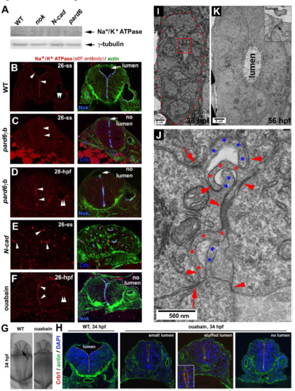
Terminal protein Na+/K+ ATPase is required to inflate the neural tube lumen, and the gut development also follows a rod-tube transition A. Western blotting revealed that Na+/K+ ATPase ?1 was expressed at 26-ss in nokm520, N-Cad, and pard6?bfh266 mutants as in wildtype. B. Na+/K+ ATPase ? co-localized with Nok at the apical surfaces in wildtype at 26-ss. C-D. Na+/K+ ATPase ? localized apically in pard6?bfh266 mutants at 28 hpf but not yet at 26-ss. E. In N-Cad mutants, Na+/K+ ATPase ? localized to the central miniature lumen of neural cellular rosettes at 26-ss. F. Ouabain treatment did not prevent Na+/K+ ATPase ? from localizing apically but did prevent the lumen from inflating. G. Dorsal view of the brain ventricles (arrow) in 34-hpf wildtype but not in ouabain-treated embryos. H. Ouabain treatment from 1-ss to 34 hpf: Crb1 and actin localized normally at the apical ends of the neural tubes (transverse sections) in wildtype (9 embryos) and in ouabain-treated embryos (30 embryos); however, ouabain-induced three defects in the lumen: small lumen (6 out of 30), stuffed lumen (stuffed with cells, inset; 4 out of 30), and no lumen (20 out of 30). I. At 36 hpf, the gut epithelial cells organized into a rod. The red-dashed line demarcates the outer boundary of the gut rod. J. A boxed region in panel I is enlarged to better show the hook-like interlocking apical membrane protrusions of gut rod cells (red and blue asterisks). These hook-like apical protrusions are presumably the microvilli, and they were adhered together with adhesions (arrowheads) to join opposing cells together. In addition, the parallel apical adhesion complexes (arrows) adhered together neighboring cells of the same orientations. K. At 56 hpf, the gut tissue developed into a hollow tube. Microvilli were no longer interlocked with each other via opposing adhesions, and a lumen emerged. However, the parallel apical adhesion persisted (inset, arrow). |

