
Fig. 3
- ID
- ZDB-FIG-180821-43
- Publication
- Fukui et al., 2018 - Hippo signaling determines the number of venous pole cells that originate from the anterior lateral plate mesoderm in zebrafish
- Other Figures
- All Figure Page
- Back to All Figure Page
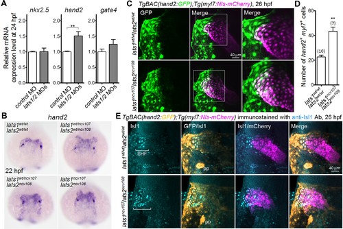
Knockout of lats1/2 results in an increase in the number of cells in the venous pole in which both myl7 and hand2 promoters are activated. (A) Quantitative-PCR analyses of the expression of nkx2.5, hand2, and gata4 mRNAs in the whole embryos at 24 hpf showing the effects of MO injection (n = 4). Relative expression of mRNA in the MO-injected morphants to that of the control is shown. (B) WISH analyses at 22 hpf of lats1wt/wtlats2wt/wt (n = 7), lats1wt/ncv107lats2wt/ncv108 (n = 22), lats1wt/ncv107lats2ncv108 (n = 14) and lats1ncv107lats2ncv108 (n = 6) embryos using an antisense probe for hand2. (C) Confocal 3D-stack images (at 26 hpf) of TgBAC(hand2:GFP);Tg(myl7:Nls-mCherry)-labeled embryos carrying the lats1wt/wtlats2wt/wt (upper panels) or lats1ncv107lats2ncv108 allele (bottom panels). GFP images (left), merged GFP and mCherry images (center), and enlarged images of the boxed regions in the center panels (right) are shown. (D) Quantitative analysis of the number of cells in which both hand2 and myl7 promoters are activated at 26 hpf. (E) Confocal 3D-stack images (at 26 hpf) of the TgBAC(hand2:GFP);Tg(myl7:Nls-mCherry)-labeled embryos containing the lats1wt/wtlats2wt/wt (upper panels, n = 9) or the lats1ncv107lats2ncv108 allele (bottom panels, n = 5) immunostained with the anti-Isl1 antibody (anti-Isl1 Ab). Square brackets denote the SHF cells that are Isl1-positive, both hand2- and myl7-promoter-active cells that are Isl1-positive, and hand2-promoter-active cells that are in contact with myl7-promoter-active cells. pp indicates the pharyngeal pouch, which expresses the hand2-promoter-activated GFP signal. The first, second, third and fourth columns show Isl1 immunostaining, merged images of GFP and Isl1 immunostaining, merged images of Isl1 immunostaining and mCherry labeling, and merged images of all the three (GFP, mCherry, and Isl1 immunostaining), respectively. All of the images are of the dorsal view, anterior to the top. The confocal 3D-stack images and the WISH images are a set of representative images from at least four independent experiments. **p < 0.01. |
| Genes: | |
|---|---|
| Antibody: | |
| Fish: | |
| Knockdown Reagents: | |
| Anatomical Terms: | |
| Stage Range: | 26+ somites to Prim-5 |
| Fish: | |
|---|---|
| Knockdown Reagents: | |
| Observed In: | |
| Stage Range: | 26+ somites to Prim-5 |