
Fig. S6
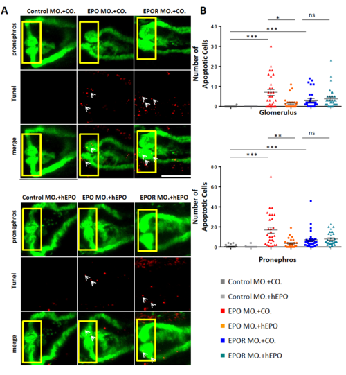
hEPO partially reduced apoptotic cell number in EPO morphants but not in EPOR morphants. A. Normalization of apoptotic cell number (white arrows) and pronephros structure in EPO morphants, but not in EPOR morphants injected with hEPO at 24 hpf. Confocal images of TUNEL-stained TG(WT1B:EGFP) zebrafish embryos at 48 hpf were shown. Arrows labelled apoptotic cells in red; pronephros was labelled in green. White scale bar: 100 ?m. B. Quantification of data for glomerulus (top) and pronephros (below) performed in three independent experiments. (n = 26?31 embryos per group). All data were analyzed using the Student's t-test. Mean � s.e.m. ns not significant. *p < 0.05. **p < 0.01. ***p < 0.001. |