
Fig. 4
- ID
- ZDB-FIG-180418-31
- Publication
- Gu et al., 2017 - Zebrafish Larvae Model of Dilated Cardiomyopathy Induced by Terfenadine
- Other Figures
- All Figure Page
- Back to All Figure Page
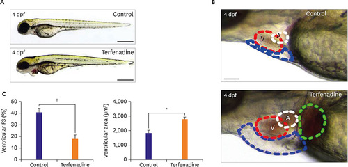
Transient terfenadine treatment impaired cardiac contraction, resulting in HF. (A) Representative images of control (0.001% DMSO) or terfenadine (20 ?M)-treated zebrafish larva. (B) Lateral view of zebrafish larvae at 4 dpf. The control zebrafish (0.001% DMSO) exhibited normal cardiac morphology, whereas terfenadine (20 ?M)-treated zebrafish larvae showed pericardial edema (blue circle) and venous congestion (green circle). (C) Quantification of ventricular FS in control (0.001% DMSO) and terfenadine (20 ?M)-treated zebrafish larvae. Ventricle size of control (0.001% DMSO) and terfenadine (20 ?M)-treated zebrafish larvae after a 24 hours treatment. n=20 zebrafish/group, scale bar=0.5 mm. a = Atrium; DMSO = dimethyl sulfoxide; dpf = days post fertilization; FS = fractional shortening; HF = heart failure; V = ventricle. *p<0.050; ?p<0.010. |
| Fish: | |
|---|---|
| Condition: | |
| Observed In: | |
| Stage: | Day 4 |