
Fig. 7
- ID
- ZDB-FIG-180410-8
- Publication
- Nagashima et al., 2017 - Anisotropic M�ller glial scaffolding supports a multiplex lattice mosaic of photoreceptors in zebrafish retina
- Other Figures
- All Figure Page
- Back to All Figure Page
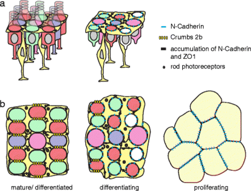
A model for how the multiplex photoreceptor lattice mosaic is patterned by anisotropic tension, glial scaffolding, and planar polarized cell adhesion. Retinal margin in a lateral and b planar views with expression and localization of cell-cell adhesion complexes (N-cadherin, Crb2b, and ZO-1) as photoreceptors and M�ller glia progress from proliferating progenitors (right) to differentiating cells in the pre-column zone (center) to the fully patterned, mature cone lattice mosaic (left). Cones (red, green, blue with UV represented by magenta); rods (small black dots); M�ller glia (bright yellow); retinal precursors (light yellow). See Discussion for additional details |