
Fig. 1
- ID
- ZDB-FIG-180208-17
- Publication
- Dudczig et al., 2017 - Developmental and adult characterization of secretagogin expressing amacrine cells in zebrafish retina
- Other Figures
- All Figure Page
- Back to All Figure Page
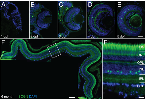
Secretagogin expression in embryonic and adult zebrafish retina. Micrographs of vertical sections through zebrafish retina immunohistochemically labeled for secretagogin (SCGN?green) with nuclei counterstained by DAPI (blue). (A?E) Sections through retinas at 1?5 days post fertilization (dpf) show earliest secretagogin positive cells detected at 3 dpf (C) and maintained at subsequent days. (F) Collage through retinal section in 6 month old zebrafish. Secretagogin expression in the amacrine layer in the inner half of the inner nuclear layer (INL) remains strong throughout adulthood. (F') Higher magnification inset of boxed region in F shows secretagogin labeled with stained processes showing monostratified band in the center of the inner plexiform layer (IPL). OS: outer segments; ONL: outer nuclear layer; OPL: outer plexiform layer; GCL: ganglion cell layer. Scale bar (E) for A-E is 50 ?m, scale bar (F) is 100 ?m, scale bar (F') is 20 ?m. |