
FIGURE
Fig. 6
- ID
- ZDB-FIG-180126-72
- Publication
- Pei et al., 2016 - Extracellular HSP60 triggers tissue regeneration and wound healing by regulating inflammation and cell proliferation
- Other Figures
- All Figure Page
- Back to All Figure Page
Fig. 6
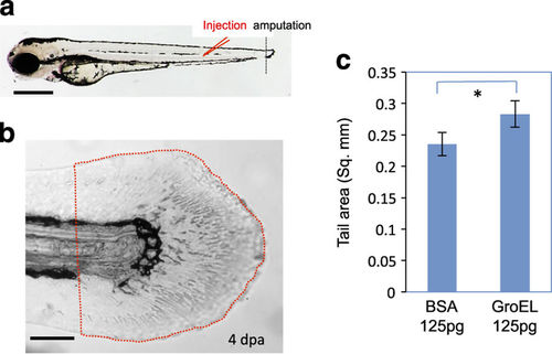
Extracellular HSP60 promotes caudal fin regeneration. (a) Schematic of injection site and amputation site. (b) Caudal fin area measured in the injected embryos at 4 dpa. Quantified areas are framed with dotted red lines, starting from the anterior end of the ventral pigmentation break. (c) Quantification of caudal fin regeneration in GroEL- and BSA-injected embryos. A significant increase (indicated by an asterisk) in the fin area is detected in GroEL-injected embryos (n=10, P=0.003). Bars = 500??m in a, 100??m in b. BSA, bovine serum albumin; dpa, day post amputation. |
Expression Data
Expression Detail
Antibody Labeling
Phenotype Data
Phenotype Detail
Acknowledgments
This image is the copyrighted work of the attributed author or publisher, and
ZFIN has permission only to display this image to its users.
Additional permissions should be obtained from the applicable author or publisher of the image.
Full text @ NPJ Regen Med