
FIGURE
Fig. 3
- ID
- ZDB-FIG-180108-2
- Publication
- Figueroa et al., 2017 - Reprimo tissue-specific expression pattern is conserved between zebrafish and human
- Other Figures
- All Figure Page
- Back to All Figure Page
Fig. 3
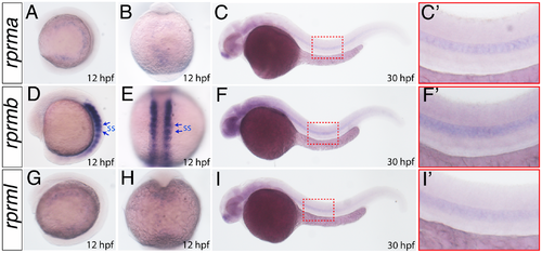
rprm expression patterns during early embryogenesis. (A-I) The expression patterns of rprma, rprmb and rprml were visualized by whole-mount in situ hybridization (WISH) during zebrafish embryonic development. Developmental stages are expressed as hours post-fertilization (hpf). (A, C, D, F, G, I) Lateral (anterior to the left) and (B, E, H) dorsal views are shown. Blue arrows in D-E indicate the expression in the somites (ss), while (C, F, I) the red inset magnifications indicate the expression in the notochord. |
Expression Data
| Genes: | |
|---|---|
| Fish: | |
| Anatomical Terms: | |
| Stage Range: | 5-9 somites to Prim-15 |
Expression Detail
Antibody Labeling
Phenotype Data
Phenotype Detail
Acknowledgments
This image is the copyrighted work of the attributed author or publisher, and
ZFIN has permission only to display this image to its users.
Additional permissions should be obtained from the applicable author or publisher of the image.
Full text @ PLoS One