
Fig. S1
- ID
- ZDB-FIG-171227-10
- Publication
- Shao et al., 2017 - Vegetally localised Vrtn functions as a novel repressor to modulate bmp2b transcription during dorsoventral patterning in zebrafish.
- Other Figures
- All Figure Page
- Back to All Figure Page
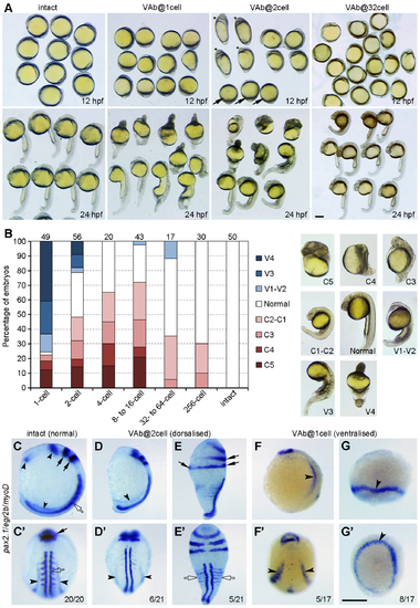
Stage-dependent dorsalising effect of VAb. (A) Representative phenotypes following VAb at indicated stages. Asterisks and arrows denote dorsalised and ventralised embryos, respectively. (B) Bar graph shows the stage-dependent dorsalising effect of VAb. Numbers on the top indicate total embryos scored from three independent experiments. Different degrees of dorsalised (C5-C1) and ventralised (V4-V1) phenotypes are shown on the right. (C-G') Dorsalised and ventralised embryos at 13 hpf revealed by simultaneous ISH. pax2.1 labels optic field, midbrain-hindbrain boundary and pronephros (black arrowheads), egr2b (krox20) labels the hindbrain (black arrows), and myoD marks the somites (open arrows). The images are shown in lateral (C,D,E,F,G) and dorsal (C',D',E',F',G') views, with anterior on the top. (C,C') An intact embryo. (D,D') A mildly dorsalised VAb@2cell embryo. (E,E') A severely dorsalised VAb@2cell embryo with radial neural marker expression and expanded paraxial tissue. (F,F') A mildly ventralised VAb@1cell embryo. (G,G') A radially ventralised VAb@1cell embryo with only pronephric progenitor expression of pax2.1. Scale bars: 250 μm. |