
Fig. 7
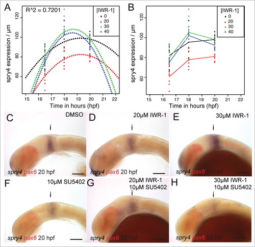
spry4 shows a bivalent response to changes in Wnt and FGF signaling. Polynomials were fitted against the spatial expression of spry4 in the dorsal midbrain (�m) at 16.5, 18, 20 hpf following treatment with IWR-1 from 14 hpf (A) and showed a good fit (R2 = 0.7201, n = 10 for each condition). A line plot of the spry4 expression domain (scaled to midbrain size) in the same embryos (B) reveals that treatment with 20�M IWR-1 had the strongest affect (n = 10 for each condition). Lateral views of 20 hpf embryos processed by in situ hybridization to reveal spry4 (blue) and pax6 (red) expression following treatment from 14 hpf with DMSO (C), 20�M IWR-1 (D), 30�M IWR-1 (E), 10�M SU5402 (F), 20�M IWR-1 with 10�M SU5402 (G), or 30�M IWR-1 with 10�M SU5402 (H). Isthmus (i). Scale bars: 100�m (C?H). |