
Fig. S2
- ID
- ZDB-FIG-171127-61
- Publication
- Takita et al., 2016 - Effects of NDRG1 family proteins on photoreceptor outer segment morphology in zebrafish
- Other Figures
- All Figure Page
- Back to All Figure Page
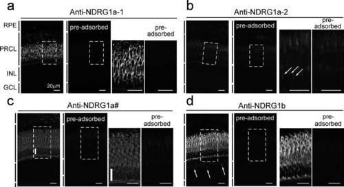
Localization of NDRG1a-1, NDRG1a-2 and NDRG1b proteins in zebrafish retina. Retinal sections were immunoprobed with anti-NDRG1a-1, anti-NDRG1a-2, anti-NDRG1a#, or anti-NDRG1b antiserum (leftmost panel in each of a - d). In (a) - (d), control measurement was made with each antiserum pre-adsorbed by each corresponding NDRG1a family protein (second left panels in a - d). The area surrounded by a dotted rectangular was magnified and is shown in the right two panels in (a) - (d). Arrows in (b) indicate the thin cone process detected by anti-NDRG1a-2 antiserum. Thick vertical white bars in (c) indicate the immunostaining of NDRG1a-1 in the plasma membranes surrounding the rod nucleus (see text). Arrows in (d) indicate NDRG1b positive cells in the inner nuclear layer. RPE, retinal pigment epithelium; PRCL, photoreceptor cell layer; INL, inner nuclear layer; GCL, ganglion cell layer. Scale bars indicate 20 ?m throughout this figure. |