Fig. S7
- ID
- ZDB-FIG-170921-47
- Publication
- Kara et al., 2017 - miR-27 regulates chondrogenesis by suppressing Focal Adhesion Kinase during pharyngeal arch development
- Other Figures
- All Figure Page
- Back to All Figure Page
|
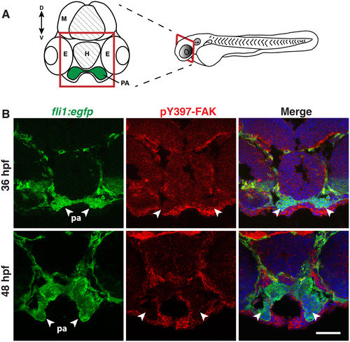
PCC cells in the pharyngeal arches have low levels of active FAK (pY397-FAK) at 36 and 48 hpf. (A) Schematic of a transverse cross-section of the zebrafish head at 36 hpf. M, mesencephalon; H, hypophysis; E, eye; PA, pharyngeal arch. Dorsal (D) is to the top, ventral (V) is to the bottom. (B) Anti-phospho FAK (pY397-FAK) and anti-GFP immunostaining on transverse sections of Tg(fli1a:eGFP)y1 embryos at 36 and 48 hpf. PCC cells in the pharyngeal arch (PA) region are indicated by arrowheads. Scale bar, 50 Ám. (C) Anti-phospho FAK (pY397-FAK), anti-phalloidin (to label cell borders) and nuclear TO-PRO-3 immunostaining on transverse sections of telencephalon in wild-type embryos at 29 hpf. Embryos were injected with either 3 ng MO-ctl or MO-ptk2 at the single cell stage. The indicated ratio represents the number of embryos with the represented phenotype/total number of observed embryos. Scale bar, 10 Ám. |
Reprinted from Developmental Biology, 429(1), Kara, N., Wei, C., Commanday, A.C., Patton, J.G., miR-27 regulates chondrogenesis by suppressing Focal Adhesion Kinase during pharyngeal arch development, 321-334, Copyright (2017) with permission from Elsevier. Full text @ Dev. Biol.

