
Fig. 3
- ID
- ZDB-FIG-170824-32
- Publication
- Helm et al., 2017 - Partial uniparental isodisomy of chromosome 16 unmasks a deleterious biallelic mutation in IFT140 that causes Mainzer-Saldino syndrome
- Other Figures
- All Figure Page
- Back to All Figure Page
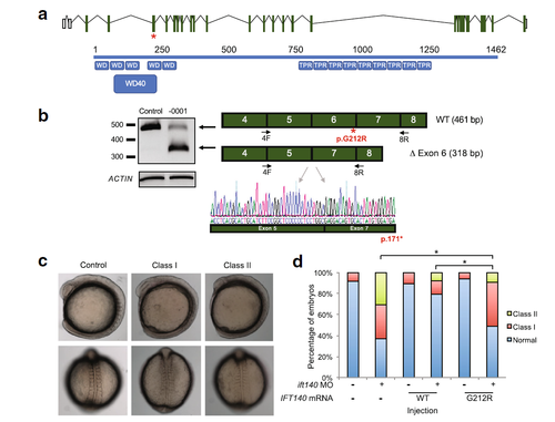
In vitro and in vivo functional analysis of IFT140 c.634G>A; p.Gly212Arg. a Schematic of the human IFT140 locus at chr16:1,510,427-1,612,110 (hg19; top) and translated protein (bottom). Exons are depicted as green boxes; untranslated regions are shown as white boxes (NM_014714.3). Protein schematic (blue; NP_055529.2) indicates predicted WD40 repeat (WD40) and tetratricopeptide-like helical domains (TPR). IFT140 c.634G > A; p.Gly212Arg location is indicated with a red star. b RT-PCR from proband lymphocyte cell lines indicating aberrant mRNA splicing. IFT140 cDNA was amplified with primers flanking the exon 6 canonical splice donor site (black arrows). Sequence chromatograms from purified PCR product indicates that the majority of IFT140 message is missing exon 6 (bottom). c Lateral (top) and dorsal (bottom) views of ift140 morphants exhibiting gastrulation defects. d In vivo complementation studies. Live embryo batches were scored to assay variant pathogenicity. Class I, modest shortening of the body axis and reduction in size of anterior structures; class II, severe shortening of the body axis and decreased anterior structures accompanied by broadening and/or kinking of the notochord and thinning of the somites. To compare pairs of embryo batches, χ 2 tests were used; p < 0.0001 indicated with (asterisk) |
| Fish: | |
|---|---|
| Knockdown Reagent: | |
| Observed In: | |
| Stage: | 5-9 somites |