
Fig. S1
- ID
- ZDB-FIG-170815-32
- Publication
- Mazon-Moya et al., 2017 - Septins restrict inflammation and protect zebrafish larvae from Shigella infection
- Other Figures
- All Figure Page
- Back to All Figure Page
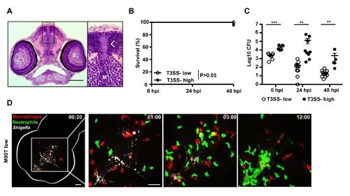
S. flexneri infection of the zebrafish hindbrain ventricle. A. Transverse section of larvae (3 dpf) infected in the HBV with S. flexneri M90T (low dose) for 6h. Arrow indicates the localisation of the bacteria inside the ventricle. Scale bar, 50 ?m. E, eye; M, midbrain ventricle. B. Survival curves of larvae infected with low (? 3 x 10³ CFU) or high (? 1 x 10⁴ CFU) inoculum of T3SS- S. flexneri (?mxiD strain) using at least 15 larvae per experiment. Significance testing performed by Log Rank test. C. Enumeration of bacteria at 0, 24, or 48 hpi from larvae infected with low (open circles) or high (closed circles) dose of S. flexneri M90T using up to 3 larvae per treatment. Circles represent individual larvae. Mean � SEM also shown (horizontal bars). Significance testing performed by Student?s t test. **, P<0.01; ***, P<0.001. Note bacterial load does not significantly decrease in highly infected fish because of the high bacteria:leukocyte ratio and thus more time is required to clear the bacterial burden. D. Frames extracted from in vivo time-lapse confocal imaging of mpeg1:G/U:mCherry x mpx:GFP larvae (3 dpf) injected in the HBV with low dose of Crimson-S. flexneri. First frame 20 mpi, followed by frames at 1, 3, and 12 hpi. Maximum intensity Z-projection images (2 ?m serial optical sections) are shown. Scale bars, 50 ?m. See also S1 Video. |