Fig. 3
- ID
- ZDB-FIG-161116-22
- Publication
- Liu et al., 2016 - A High-Content Larval Zebrafish Brain Imaging Method for Small Molecule Drug Discovery
- Other Figures
- All Figure Page
- Back to All Figure Page
|
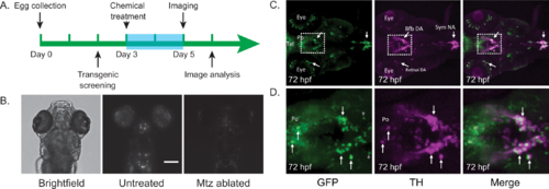
Chemo-genetic model screening details. (A) Timeline showing the details of fish husbandry and chemical treatment. Larvae were treated with metronidazole at 3dpf and imaged in situ at 5dpf. (B) The nitroreductase-metronidazole method is able to specifically ablate dopaminergic neurons, which express mCherry, in 5dpf larvae (scale bar = 200um). Images are of zebrafish larvae in the dorsal-down position. (C) Anti-tyrosine hydroxylase and anti-GFP antibody staining of 3dpf zebrafish embryos show good overlap in the ventral forebrain region DA neurons. bfb DA, basal forebrain dopaminergic neurons; Po, preoptic region; sym NA, sympathetic noradrenergic neurons; Tel, telencephalon; retinal DA, retinal dopaminergic neurons. (D) Zoomed-in views of areas boxed in (C). |

