
Fig. S1
- ID
- ZDB-FIG-160926-28
- Publication
- Monteiro et al., 2016 - Transforming Growth Factor ? Drives Hemogenic Endothelium Programming and the Transition to Hematopoietic Stem Cells
- Other Figures
- All Figure Page
- Back to All Figure Page
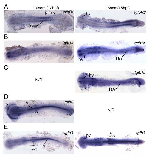
Expression of TGFβ signalling components at 12hpf (10somite stage) and 15hpf (16somite stage). All embryos were flatmounted for imaging. (A) Expression of tgfbR2 in the somites (som) and posterior lateral mesoderm (plm) at 12hpf. Expression of tgfbR2 at 15hpf in head vasculature (hv) and somites. (B) tgfb1a is not expressed in the posterior at 12hpf but is present in head vasculature from 12hpf and in the embryonic dorsal aorta (DA) at 15hpf. (C) tgfb1b is also present in the head vasculture and DA at 15hpf. Note that there is anterior expression of tgfb1b in cells that are likely myeloid. (D) tgfb2 is expressed in the polster (p), otic vesicles (o), notochord (n) and in the tailbud (tb) at 12hpf. (E) Expression of tgfb3 in the notochord and in the 4 anteriormost somites (ant som) at 12hpf. At 15hpf, the expression in the somites and notchord is maintained and weak expression in the head vasculature is observed. |
| Genes: | |
|---|---|
| Fish: | |
| Anatomical Terms: | |
| Stage Range: | 10-13 somites to 14-19 somites |
Reprinted from Developmental Cell, 38(4), Monteiro, R., Pinheiro, P., Joseph, N., Peterkin, T., Koth, J., Repapi, E., Bonkhofer, F., Kirmizitas, A., Patient, R., Transforming Growth Factor ? Drives Hemogenic Endothelium Programming and the Transition to Hematopoietic Stem Cells, 358-70, Copyright (2016) with permission from Elsevier. Full text @ Dev. Cell