
Fig. 5
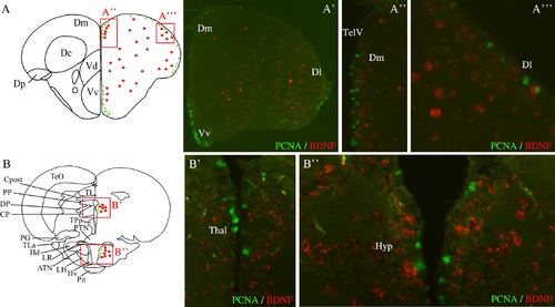
Immunohistochemical characterization of bdnf-expressing cells in adult zebrafish brain. Double staining for bdnf mRNA (red) and PCNA protein (green) on cross-sections through the telencephalon (A to A′′′), the thalamus (B and B′) and the ventral hypothalamus (B and B′′). A and B are representative sections taken from the zebrafish atlas (Wullimann et al., 1996). Bdnf-expressing cells are represented by red dots and PCNA-labeled cells are green dots. Dl: lateral zone of the dorsal telencephalon; Dm: medial zone of the dorsal telencephalon; Hyp: hypothalamus; TelV: telencephalic ventricle; Thal: thalamus; Vv: ventral zone of the ventral telencephalon. Scale bar = 200 µm in A′; 100 µm in A′′; 50 µm in A′′′. |