
Fig. S5
- ID
- ZDB-FIG-160606-22
- Publication
- Johnson et al., 2016 - Gfap-positive radial glial cells are an essential progenitor population for later-born neurons and glia in the zebrafish spinal cord
- Other Figures
- All Figure Page
- Back to All Figure Page
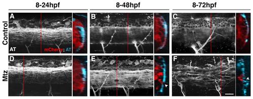
Loss of radial glia disrupts spinal cord axon organization (A-F) Lateral views of spinal cord axons within control and Mtz treated gfap:nfsbmcherry sc059 embryos immunolabeled with anti-acetylated tubulin (AT, white). Alongside each image is an excerpt from the Z stack rotated along the X-axis to show correlations between NTRmCherry (red) and AT labeling (blue). Red line depicts the location along the Z-axis where the rotated optical section is found. (A-C) Uniform, uninterrupted axon tracts are found throughout the spinal cords of control treated embryos at all time points assayed. (D) At 24hpf, no observable difference is seen (A). (E) At the onset of ablation, defects in axon organization can be observed in the spinal cord. When rotated, these defects (arrowheads) align with regions with reduced NTR-mCherry labeling or NTR-mCherry+ debris, which becomes more severe over time (F). No radial glial or axonal defects were observed in Mtz-treated gfap:eGFPmi2001 embryos at any ages assayed throughout this study (data not shown), demonstrating that any observed phenotypes correspond to the direct loss of radial glia. Scale bar = 20�m. |