
Fig. 6
- ID
- ZDB-FIG-160311-49
- Publication
- Welker et al., 2016 - Standardized orthotopic xenografts in zebrafish reveal glioma cell line specific characteristics and tumor cell heterogeneity
- Other Figures
- All Figure Page
- Back to All Figure Page
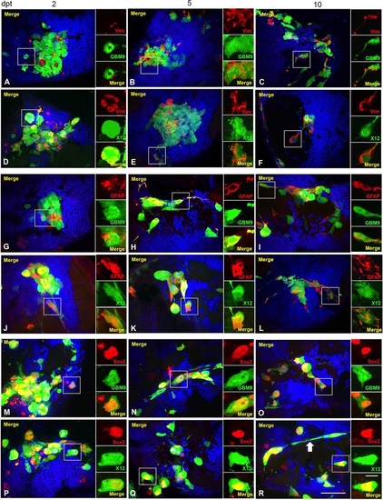
GBM9 and X12 tumors contain a combination of differentiated cells and stem cells. Confocal images of GBM9 and X12 on 2 (A,D,G,J,M,P), 5 (B,E,H,K,N,Q) and 10 (C,F,I,L,O,R) dpt transverse cryosections. (A-C) GBM9 (green), DAPI (blue) and vimentin (red) at 100�. (D-F) X12 (green), DAPI (blue) and vimentin (red) at 100�. (G-I) GBM9 (green), DAPI (blue) and GFAP (red) at 100�. (J-L) X12 (green), DAPI (blue) and GFAP (red) at 100�. (M-O) GBM9 (green), DAPI (blue) and Sox2 (red) at 100�. (P-R) X12 (green), DAPI (blue) and Sox2 (red) at 100�. White boxes denote area magnified to the right of the image. White arrow in R points to a cell with a migratory morphology. n=5 animals per group; 90 total animals. Scale bar: 20�m for main panels and 5�m for insets. |