
Fig. 6
- ID
- ZDB-FIG-151228-16
- Publication
- Mendieta-Serrano et al., 2015 - Spatial and temporal expression of zebrafish glutathione peroxidase 4 a and b genes during early embryo development
- Other Figures
- All Figure Page
- Back to All Figure Page
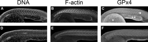
GPx4 immunofluorescence localization patterns in zebrafish embryos at 24 hpf as detected by laser confocal microscopy. Hoechst-stained embryos (A and D). F-actin, phalloidin Alexa 488-stained embryos (B and E). GPx4 immunolocalization (C and F). Embryos by 24 h of development (A, B and C) show GPx4b immunolocalization at the myotomes and at the longitudinal myosepta (C, arrow head). At slightly more advance stages (D, E and F) notice that the yolk cell extension (double asterisk) is of longer length and is narrower compared to younger embryos shown in A, B and C and show an increase in the immunlocalization signal apparently corresponding to GPx4a. The GPx4b immunolocalization is still found at the myotomes but is no longer found at the longitudinal myosepta (F). *, yolk cell. Scale bar 500 µm. |
| Antibody: | |
|---|---|
| Fish: | |
| Anatomical Terms: | |
| Stage: | Prim-5 |
Reprinted from Gene expression patterns : GEP, 19(1-2), Mendieta-Serrano, M.A., Schnabel-Peraza, D., Lomelí, H., Salas-Vidal, E., Spatial and temporal expression of zebrafish glutathione peroxidase 4 a and b genes during early embryo development, 98-107, Copyright (2015) with permission from Elsevier. Full text @ Gene Expr. Patterns