Fig. 3
- ID
- ZDB-FIG-151214-70
- Publication
- Askary et al., 2015 - Iroquois Proteins Promote Skeletal Joint Formation by Maintaining Chondrocytes in an Immature State
- Other Figures
- All Figure Page
- Back to All Figure Page
|
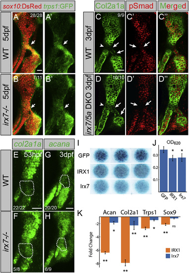
Irx7 Represses Chondrocyte Maturation (A and B) trps1:GFP (green) is reduced at the hyoid joint (arrows) of irx7el538 mutants compared with WT siblings. sox10:dsRed labels hyoid cartilages (red). (C and D) Immunostaining shows increased Col2a1a protein (green) but no change in phospho-Smad1/5/8 (red) in the hyoid joints (arrows) and Hm-Sy connecting zone (arrowheads) of irx7el538; irx5ael574 mutants relative to WT siblings. (E?H) In situ hybridization shows increased expression of col2a1a and acana in the hyoid joint region (dashed areas) of irx7el538 mutants relative to WT siblings. Numbers indicate proportion of animals showing the displayed phenotype. Scale bars represent 20 ÁM. (I) Irx7 and murine IRX1 (but not GFP control) inhibit Alcian+ matrix accumulation in micromass cultures of ATDC5 cells grown in chondrogenic media for 7 days. Results were consistent across biological triplicates. (J) Alcian blue content of the micromasses quantified by absorbance at 620 nm. (K) qRT-PCR on RNA extracted from micromass cultures of ATDC5 cells grown in chondrogenic media for 7 days. Compared with GFP-expressing controls, expression of Col2a1, Acan, and Trps1 was reduced by IRX1 and Irx7 misexpression. Sox9 expression was reduced by IRX1 but not Irx7 misexpression. Error bars represent 95% confidence interval of the mean. p < 0.05 and p < 0.01 using Tukey HSD test. See also Figure S3. |
| Genes: | |
|---|---|
| Antibodies: | |
| Fish: | |
| Anatomical Terms: | |
| Stage Range: | Long-pec to Day 5 |
Reprinted from Developmental Cell, 35, Askary, A., Mork, L., Paul, S., He, X., Izuhara, A.K., Gopalakrishnan, S., Ichida, J.K., McMahon, A.P., Dabizljevic, S., Dale, R., Mariani, F.V., Crump, J.G., Iroquois Proteins Promote Skeletal Joint Formation by Maintaining Chondrocytes in an Immature State, 358-65, Copyright (2015) with permission from Elsevier. Full text @ Dev. Cell

