
Fig. S2
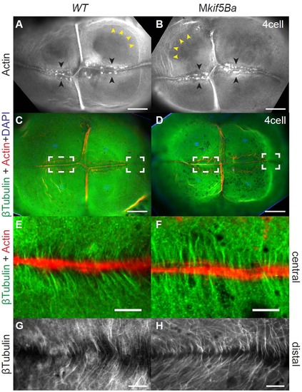
Cleavage furrow-associated cytoskeleton is intact in Mkif5Ba mutants. (A-B) Rhodamine-phalloidin staining to label F-actin. Animal pole views reveal an Factin contractile band present in all four furrows, F-actin accumulations flanking the FMA (black arrowheads) and circumferential actin bands around the blastomeres (yellow arrowheads) in both WT (A) and Mkif5Ba mutants (B). Scale bars, 100�m. (C-D) β-Tubulin antibody staining to reveal microtubules. Animal pole views show the Factin contractile band present in all four furrows and the furrow microtubule arrays that flank the newly forming furrows of WT (C) and Mkif5Ba mutants (D). Left and right dotted boxes in each panel indicate central and distal regions of the newly forming furrow respectively. Scale bars, 100�m. (E-F) Higher magnification of the central furrow shows that the furrow microtubules are parallel to each other and perpendicular to the F-actin contractile band in both WT (E) and Mkif5Ba mutants (F). Scale bars, 10�m. (G-H) Higher magnification of the distal furrow shows microtubules angled in a Vshaped configuration pointing towards the distal end in both WT (G) and Mkif5Ba mutants (H). Scale bars, 10�m. (All results are from n=11 embryos for Mkif5Baae11/ae12, n=9 for Mkif5Baae11/ae11, and n=15 for WT). |