
Fig. 2
- ID
- ZDB-FIG-151022-30
- Publication
- Gonzaga-Jauregui et al., 2015 - Exome Sequence Analysis Suggests that Genetic Burden Contributes to Phenotypic Variability and Complex Neuropathy
- Other Figures
- All Figure Page
- Back to All Figure Page
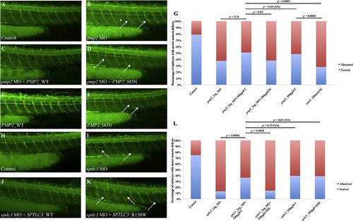
Suppression of pmp2 and sptlc3 in Zebrafish Causes Defects in Motor Axon Pathfinding and Outgrowth (A?F) Lateral views of a control embryo, an embryo injected with pmp2 morpholino (MO) and embryos injected with pmp2 MO+PMP2_WT and pmp2 MO+PMP2_I43N, PMP2_WT and PMP2_I43N cocktails, respectively, at 2 dpf (days post fertilization). Controls showed even spacing and normal branching of the motor neuron axons (A). In the pmp2 MO-injected embryos, the spacing of neuronal axons is perturbed by exiting the periphery but failing to extend (asterisks) or presenting pathfinding errors (arrows; B). Co-injection of pmp2 MO with human PMP2_WT resulted in restoration of the normal neuronal phenotype (C), but PMP2_I43N did not (D). Overexpression of human PMP2_WT causes mild pathfinding errors (E), suggesting dose sensitivity for PMP2. However, the human PMP2 mutant p.I43N was significantly more severe than PMP2_WT when overexpressed (F) and had similar effects to suppression of pmp2 by MO knockdown. (G) Percentage of normal versus abnormal embryos under the conditions being evaluated above. (H?K) Wild-type embryos (H) and sptlc3 morphants (I) in which secondary axons fail to migrate appropriately (white arrows). The phenotype induced by suppression of sptlc3 could be rescued by co-injection with SPTLC3_WT (J) but not SPTLC3_R150W (K). (L) Quantification of normal embryos versus embryos with motor neuron axon defects. For statistical analyses, ?2 tests were performed. |
| Antibody: | |
|---|---|
| Fish: | |
| Knockdown Reagents: | |
| Anatomical Terms: | |
| Stage: | Long-pec |
| Fish: | |
|---|---|
| Knockdown Reagents: | |
| Observed In: | |
| Stage: | Long-pec |