
Fig. 5
- ID
- ZDB-FIG-150923-12
- Publication
- Yan et al., 2015 - Stimulation of hepatocarcinogenesis by neutrophils upon induction of oncogenic kras expression in transgenic zebrafish
- Other Figures
- All Figure Page
- Back to All Figure Page
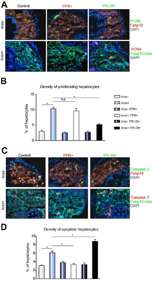
Proliferation and apoptosis analyses of effects of neutrophils on oncogenic hepatocytes. 8 dpf fabp10+ and kras+ larvae after exposure to FPR A14 or PR-39 with doxycycline were analysed by immunohistochemistry. (A and C) PCNA (A) or active caspase 3 (B) staining with representative images from each group. Control Fabp10+ larvae with DsRed expression in hepatocytes were stained with Alexa Fluor 488-conjugated secondary antibody after the primary antibody incubation (top rows) while kras+ larvae with GFP expression in hepatocytes were stained with Alexa Fluor 568-conjugated secondary antibody (bottom rows). All sections were counter-stained with DAPI. (B and D) Quantification of proliferating (B) and apoptotic (D) hepatocytes as percentage of total hepatocytes (n = 10; *p <0.05; n.s., no significance). |
| Genes: | |
|---|---|
| Antibodies: | |
| Fish: | |
| Conditions: | |
| Anatomical Term: | |
| Stage: | Days 7-13 |
| Fish: | |
|---|---|
| Conditions: | |
| Observed In: | |
| Stage: | Days 7-13 |
Reprinted from Journal of hepatology, 63(2), Yan, C., Huo, X., Wang, S., Feng, Y., Gong, Z., Stimulation of hepatocarcinogenesis by neutrophils upon induction of oncogenic kras expression in transgenic zebrafish, 420-8, Copyright (2015) with permission from Elsevier. Full text @ J. Hepatol.