
Fig. S1
- ID
- ZDB-FIG-150910-8
- Publication
- Fortuna et al., 2015 - Vascular Mural Cells Promote Noradrenergic Differentiation of Embryonic Sympathetic Neurons
- Other Figures
- All Figure Page
- Back to All Figure Page
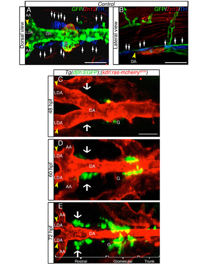
DβH-positive cells develop in proximity to the DA in live zebrafish embryos, related to Figure 1. (A, B) TH+ expressing cells are dispersed as several clusters between the LDA (arrowhead) - DA connection and the glomerular region (G). Dorsal (A) and lateral (B) views (anterior is to the left) of whole-mount 60hpf Tg(kdrl:EGFP)la116 zebrafish embryos. Tg(kdrl:EGFP)la116 zebrafish embryos immunostained with antibodies to detect GFP (vessels, green), ZN12 (glomerulus, red) and Tyrosine Hydroxylase enzyme (TH) (catecholaminergic marker, blue). (C-E) Live confocal fluorescence images (dorsal view) of Tg(dβh:EGFP);(kdrl:ras-mcherry)s916 embryos between the LDA (arrowhead) - DA connection and the glomerular region (G) at indicated stages. Anterior is to the left. (C) DβH-positive cells (green) are first detected at 48 hpf. (D) Between 48 - 60 hpf, DβH-positive cells develop in loco (white arrows) and close to the LDA-DA connection, while a new vessel (AA) is formed with LDA. (E) EGFPpositive cells are organized in three clusters (rostral, glomerular and trunk) around DA. The number of DβH-positive cells increase both rostrally and caudally along DA from 48 to 72 hpf. Data were calculated from three independent experiments. N.S., not significant; error bars indicate SD. DA, dorsal aorta; LDA, lateral dorsal aorta; AA, aortic arch; G, glomerulus; hpf, hours postfertilization. Scale bar: 75 µm in A; 50 µm in B-E. |