
FIGURE
Fig. 4
- ID
- ZDB-FIG-150602-18
- Publication
- Chang et al., 2014 - Perturbing the developing skull: using laser ablation to investigate the robustness of the infraorbital bones in zebrafish ( Danio rerio )
- Other Figures
- All Figure Page
- Back to All Figure Page
Fig. 4
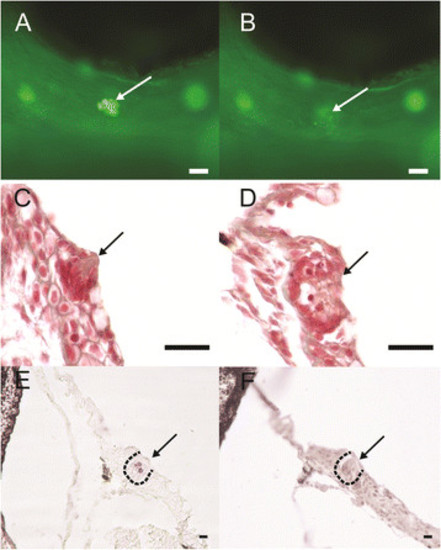
Representative figures of targeted neuromast laser ablation on infraorbital three canal neuromast. (A) Before and (B) after neuromast laser ablation in live zebrafish specimens stained with FM1-43. Masson’s trichrome stained cross-section of neuromast of (C) control-side and (D) post-ablation neuromasts. (E and F) TUNEL stained cross-section of ablated neuromasts (E) and control side (F). Arrow indicates neuromasts. Scale bars are (A and B) 25 µm and (C-F) 10 µm. |
Expression Data
Expression Detail
Antibody Labeling
Phenotype Data
Phenotype Detail
Acknowledgments
This image is the copyrighted work of the attributed author or publisher, and
ZFIN has permission only to display this image to its users.
Additional permissions should be obtained from the applicable author or publisher of the image.
Full text @ BMC Dev. Biol.