
Fig. S1
- ID
- ZDB-FIG-150430-17
- Publication
- Luz et al., 2014 - Dynamic Association with Donor Cell Filopodia and Lipid-Modification Are Essential Features of Wnt8a during Patterning of the Zebrafish Neuroectoderm
- Other Figures
- All Figure Page
- Back to All Figure Page
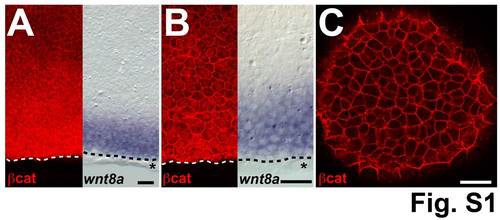
β-catenin protein has graded expression in the blastoderm margin. Comparison of β-catenin staining and wnt8a ISH of shield stage embryos. (A) β-catenin staining intensity decreases from the margin towards the animal pole (left) but is not restricted to the wnt8a mRNA expression domain (right). (B) High magnification shows nuclear β-catenin staining at the margin and decreasing intensity from the margin towards the animal pole (left) but clearly detectable anteriorly (towards the animal pole) to the wnt8a mRNA expression domain (right). (C) β-catenin staining in the animal pole is restricted to the membrane. Dashed line indicates the blastoderm margin, * indicates a row of EVL cells. A and B are representative images of different embryos, at 90� from the shield. Scale bars: 50 �m. |