
FIGURE
Fig. 1
- ID
- ZDB-FIG-150422-60
- Publication
- Olena et al., 2015 - miR-216a regulates snx5, a novel notch signaling pathway component, during zebrafish retinal development
- Other Figures
- All Figure Page
- Back to All Figure Page
Fig. 1
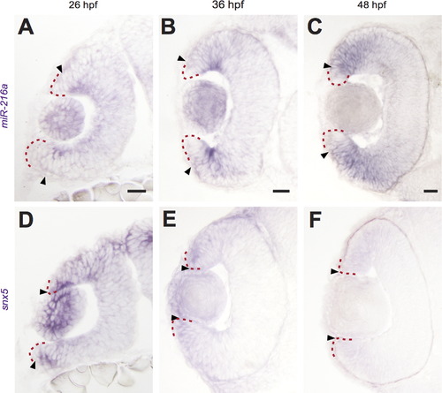
miR-216a and snx5 have complementary expression patterns during development. Transverse sections of whole mount in situ hybridizations for miR-216a and snx5 at 26 (A, D), 36 (B, E), and 48 h (C, F) post-fertilization (hpf). miR-216a expression spreads from the center of the developing retina toward the periphery. snx5 is detected in a complementary pattern becoming increasingly restricted over time to a small number of cells at the far periphery of the developing retina. Arrowheads indicate the extent of signal, the red dashed line indicates the lateral edge of the optic cup. Scale bar: 20�m. |
Expression Data
| Gene: | |
|---|---|
| Fish: | |
| Anatomical Terms: | |
| Stage Range: | Prim-5 to Long-pec |
Expression Detail
Antibody Labeling
Phenotype Data
Phenotype Detail
Acknowledgments
This image is the copyrighted work of the attributed author or publisher, and
ZFIN has permission only to display this image to its users.
Additional permissions should be obtained from the applicable author or publisher of the image.
Reprinted from Developmental Biology, 400(1), Olena, A.F., Rao, M.B., Thatcher, E.J., Wu, S.Y., Patton, J.G., miR-216a regulates snx5, a novel notch signaling pathway component, during zebrafish retinal development, 72-81, Copyright (2015) with permission from Elsevier. Full text @ Dev. Biol.