
Fig. 2
- ID
- ZDB-FIG-150331-29
- Publication
- Phng et al., 2015 - Formin-mediated actin polymerization at endothelial junctions is required for vessel lumen formation and stabilization
- Other Figures
- All Figure Page
- Back to All Figure Page
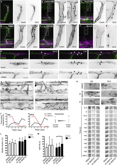
Formin Activity Promotes F-Actin Polymerization at EC Junctions (A) Coexpression of fmnl3-EGFP and DCX-mCherry in ISV at 32 hpf. Scale bar represents 10 �m. (B and C) ISVs of Tg(fli1ep:DCX-EGFP);Tg(kdr-l:ras-Cherry)s916 embryos injected with control or fmnl3 morpholino at 49 hpf. (D?F) Tg(fli1ep:DCX-EGFP);Tg(kdr-l:ras-Cherry)s916 embryos were treated with DMSO, 10 �M SMIFH2, or 0.5 �g/ml nocodazole at 48 hpf for 2 hr and imaged at 51?52 hpf. Arrowheads show microtubule filaments. Black arrows show apical membrane, and red arrows show microtubule organizing center. Scale bars represent 20 �m. (G) Mosaic endothelial fmnl3-mCherry expression in Tg(fli1ep:Lifeact-EGFP) embryo from 33 hpf. Arrowheads show localization of fmnl3 with F-actin at cell junctions. Scale bar represents 20 �m. (H?J) 3 to 4 dpf Tg(fli1ep:Lifeact-EGFP) embryos were treated with DMSO or 10 �M SMIFH2 for 4?5 hr. Arrowheads show serrated F-actin cables. DA, dorsal aorta. PCV, posterior cardinal vein. SIA, subintestinal artery. Scale bars represent 20�m. (J) Image analysis of F-actin cable profile at junctions of the DA or PCV. (K?M) Fluorescence recovery of EGFP-Actin at cell junctions after photobleaching. Three dpf Tg(fli1ep:EGFP-Actin) embryos were treated with DMSO for 5 hr or 10 �M SMIFH for 3 or 5 hr prior to photobleaching. Scale bar represents 10 �m. Plots of EGFP-Actin mobile fraction (L) and half-life (M) are shown from different treatments. Data represent mean � SD. See also Figure S2. |
Reprinted from Developmental Cell, 32, Phng, L.K., Gebala, V., Bentley, K., Philippides, A., Wacker, A., Mathivet, T., Sauteur, L., Stanchi, F., Belting, H.G., Affolter, M., Gerhardt, H., Formin-mediated actin polymerization at endothelial junctions is required for vessel lumen formation and stabilization, 123-32, Copyright (2015) with permission from Elsevier. Full text @ Dev. Cell