
Fig. 7
- ID
- ZDB-FIG-150312-45
- Publication
- Feng et al., 2014 - Negative feedback regulation of Wnt signaling via N-linked fucosylation in zebrafish
- Other Figures
- All Figure Page
- Back to All Figure Page
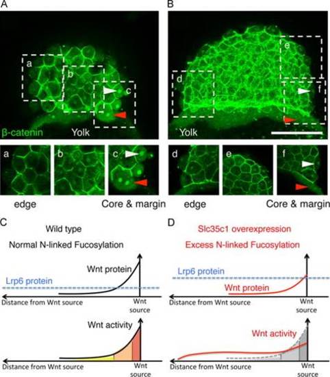
Excess fucosylation changes the response pattern of Wnt signaling. (A-B) Projections of stacks of individual confocal image sections show the nuclear localization of β-catenin in early zebrafish embryos (512 cell stage). (a?f) Single confocal sections. (A) β-catenin in a representative wild-type embryo. (a) Peripheral blastomeres at the animal pole. (b) medial region of the blastoderm. (c) The ?core? region of highest Wnt activity at the dorsal margin. (B) The Wnt gradient of a representative mSlc35c1 expressing embryo. (d) The edge, (e) the medial and (f) the core and margin regions. (C-D) Schematic illustration shows Wnt activities based on β-catenin nuclear localization. In wild-type embryos, with the normal level of N-fucosylation, Wnt protein creates a gradient. In Slc35c1 over-expressing embryos, with excess N-fucosylation, Wnt protein levels are diminished, which reduces the highest Wnt activity response region; Lrp6 protein level is increased, which increases the basal level of the Wnt response, creating an extended zone of intermediate Wnt response. Note: Bar=200 �m. |
| Antibody: | |
|---|---|
| Fish: | |
| Anatomical Term: | |
| Stage: | 512-cell |
Reprinted from Developmental Biology, 395(2), Feng, L., Jiang, H., Wu, P., Marlow, F.L., Negative feedback regulation of Wnt signaling via N-linked fucosylation in zebrafish, 268-86, Copyright (2014) with permission from Elsevier. Full text @ Dev. Biol.