Fig. 1
- ID
- ZDB-FIG-150121-13
- Publication
- Wanglar et al., 2014 - Tbx Protein Level Critical for Clock-Mediated Somite Positioning Is Regulated through Interaction between Tbx and Ripply
- Other Figures
- All Figure Page
- Back to All Figure Page
|
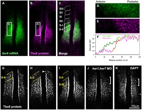
Periodic expression of Tbx6 protein and posttranscriptional regulation of its anterior border. (A-C) In situ hybridization with tbx6 probe (A) and immunostaining with anti-Tbx6 antibody (B) were performed using zebrafish embryos at the 8 somite stage (n = 15). Merged image (C) combined (A) and (B) is also shown. Zebrafish tbx6 mRNA is expressed broadly throughout the anterior PSM. At the same time, the Tbx6 protein is also expressed throughout the anterior PSM, however, its anterior border is restricted far posterior to the anterior border of the mRNA. The position of each segmental unit is also indicated from SIII to S-II. (D-F) Quantitative analysis of tbx6 mRNA and protein in the PSM. Intensity of tbx6 mRNA signals in a boxed area in the PSM (D; the boxed area shown in (A) is indicated by 90° rotation) and protein signals in the corresponding area (E; the boxed area shown in (B) is indicated by 90° rotation) was scanned and indicated by green and magenta lines, respectively in (F). While tbx6 mRNA is gradually decreased in the anterior region (shown by dark green line), Tbx6 protein level is abruptly decreased (shown by red line). Anterior is left and posterior is right. (G-I) Indication of 3 typical patterns of embryos stained with anti-Tbx6 antibody. Embryos were observed at 8 somite stage. Comparative analysis with her1 mRNA expression shown in Figure 2 indicates that the anterior border of the Tbx6 protein follows a phase of periodic change. After a long core domain of Tbx6 proteins is generated (G), an anterior part of the Tbx6 protein domain was eliminated, resulting in appearance of the upper band, which is indicated by an arrowhead (H), then this upper band disappeared, resulting in a short Tbx6 domain (I). Out of a total of 154 embryos examined, around 40% of them showed (G), 35% showed (H), 25% showed (I) type of expression pattern. (J) A 10 somite stage embryo injected with both her1 and her7 specific antisense morpholino oligos was stained with anti-Tbx6 antibody. The defects were observed in 97.5% of the injected embryos (n = 40). (K) A 10 somite stage embryo treated with DAPT, a Notch inhibitor, was stained with anti-Tbx6 antibody. The defects were observed in all of the embryos treated with DAPT (n = 22). Pattern of Tbx6 proteins was disturbed in anterior area indicated by a bracket (J, K). The yellow dotted lines indicate S-II (G, H) and S-II and S-III (I) regions. |
| Gene: | |
|---|---|
| Antibody: | |
| Fish: | |
| Condition: | |
| Knockdown Reagents: | |
| Anatomical Term: | |
| Stage Range: | 5-9 somites to 10-13 somites |

