
Fig. 4
- ID
- ZDB-FIG-141016-43
- Publication
- Hudish et al., 2013 - miR-219 Regulates Neural Precursor Differentiation by Direct Inhibition of Apical Par Polarity Proteins
- Other Figures
- All Figure Page
- Back to All Figure Page
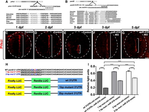
miR-219 Has Single, Conserved Target Sites within prkci and pard3 32 UTRs (A and B) Schematic representations of prkci and pard3 transcripts with predicted miR-219 target sites conserved among various species. (C?E) In controls at 1 and 2 dpf, Prkci protein is concentrated at apical membranes lining the primitive lumen, but by 3 dpf Prkci is limited to the central canal (brackets). (F and G) Prkci labeling persists along a primitive lumen extending across the spinal cord dorsoventral axis in 3 dpf and 5 dpf miR-219 MO-injected larvae. (H) Sequences (220 bp) from the pard3 32 UTR containing wild-type and mutated miR-219 target sites were cloned into dual luciferase vectors. (I) Quantification of light units revealed a miR-219-mediated reduction of reporter gene expression that was abrogated by 1 and 2 bp mutations within the target site. Data represent � SEM (three independent experiments). Brackets indicate pairwise comparisons. p < 0.0001, unpaired t test. Scale bar equals 10 μm. |
| Antibody: | |
|---|---|
| Fish: | |
| Knockdown Reagent: | |
| Anatomical Terms: | |
| Stage Range: | Prim-5 to Day 5 |
| Fish: | |
|---|---|
| Knockdown Reagent: | |
| Observed In: | |
| Stage Range: | Protruding-mouth to Day 5 |
Reprinted from Developmental Cell, 27(4), Hudish, L.I., Blasky, A.J., and Appel, B., miR-219 Regulates Neural Precursor Differentiation by Direct Inhibition of Apical Par Polarity Proteins, 387-398, Copyright (2013) with permission from Elsevier. Full text @ Dev. Cell