Fig. 1
- ID
- ZDB-FIG-140903-8
- Publication
- Heude et al., 2014 - The dlx5a/dlx6a Genes Play Essential Roles in the Early Development of Zebrafish Median Fin and Pectoral Structures
- Other Figures
- All Figure Page
- Back to All Figure Page
|
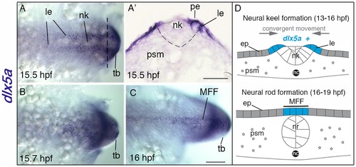
Expression of dlx5a during early specification of median fin fold ectodermal cells. (A?C) In situ hybridization for dlx5a in zebrafish embryos from 15.5 hpf to 16 hpf: dorsal view of the posterior axis (A, C) and coronal section (A2) at the level indicated by the dashed line in (A). At 15.5 hpf, dlx5a is expressed in ectodermal cells at the lateral edges (le) of the neural keel (nk) underlying the periderm (pe) (A, A2) (the dashed line in A2 delineates the neural keel). From 15.5 hpf to 16 hpf, ectodermal cells expressing dlx5a follow a dynamic convergent movement to form the presumptive median fin fold (MFF) at the dorsal midline of the embryo (B?C). (D) Schematic representation of the zebrafish dorsal cellular movement implicating dlx5a based on A?C. The convergent movement produced by the establishment of the neural rod (nr) (16?19 hpf) leads to the fusion of the two lateral edges at the midline into the presumptive MFF expressing dlx5a. ep, epidermis; nc, notochord; psm, presomitic mesoderm; tb, tail bud. Scale bars shown in C for A?C and in A2 50 μm. |
| Gene: | |
|---|---|
| Fish: | |
| Anatomical Terms: | |
| Stage Range: | 10-13 somites to 14-19 somites |

