
Fig. S8
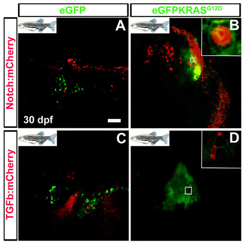
KRAS positive cells expressing Notch and TGFβ signaling reporters during MDB progression. Confocal zoom was performed on cerebellum tissue to observe the activity of TGFβ and Notch pathways at single-cell level. Normal (A and C) and Hyperplastic cerebella (B and D) are depicted, with scale bars for original (50 μm; A, B, C, D). The evidenced enlargement in panel B shows KRAS dependent activity of TGFβ pathway in MDB (tumor-prone green cells are also red reporter-positive). The enlargement in panel D shows KRAS dependent activity of Notch pathway in MDB (tumor-prone green cells are also red reporter-positive). Both pathways appear simultaneously involved in different ways during MDB onset and progression. |