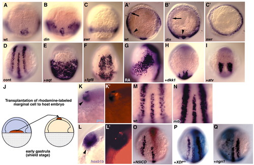
Regulation of pnx expression. (A-C2) Expression of pnx in wild-type (A,A2), chordino (B,B2) and swirl (C,C2) mutant embryos at the mid-gastrula stage (85% epiboly). (A-C) Dorsal views. (A2-C2) Vegetal pole views, with dorsal towards the bottom. Arrows and arrowheads indicate the ventral and dorsal limit of lateral expression domains of pnx, respectively. Lateral expression domains of pnx, which correspond to the primary inter neurons and Rohon-Beard (RB) neurons, became closer to the dorsal midline in chordino mutant embryos. By contrast, the pnx transcripts were detected in the entire ectoderm margin at the mid gastrula stage in swirl mutant embryos. (D-I) Reciprocal regulation of pnx by prospective posteriorizing and anteriorizing signals. The expression of pnx was expanded or ectopically induced at the three-somite stage in the embryos injected with 0.5 pg of sqt RNA (E) and 10 pg of fgf8/ace RNA (F), or treated with 1 μM retinoic acid at the gastrula stage (G) (D, control). (D-F) Dorsal views and (G) lateral view. (H,I) pnx expression was reduced and the expression domain was posteriorly shifted in embryos injected with 20 pg of dkk1 RNA (H) and 1 pg of antivin (I) RNA at the three-somite stage. Dorsal views. (J-L2) Non-axial mesendoderm induced the ectopic expression of pnx and hoxb1b in the forebrain region. (J) Procedure of the experiment. Marginal blastomere cells from the rhodamine-dextran-injected embryos were transplanted into the animal-pole region of host embryos at the shield stage, which were then stained at the three-somite-stage for pnx (K,K2) or hoxb1b (L,L2). Lateral views, with dorsal towards the right (K-L2). (K2,L2) Epifluorescence images of K,L, respectively. Ectopic expression of pnx and hoxb1b is indicated by arrowheads. (M-Q) Regulation of pnx by the Delta-Notch signal. The number of pnx-expressing cells at the three-somite stage in mind bomb (mib) mutant embryos (N) and embryos injected with RNA of 100 pg of XDlstu RNA (P) was increased in comparison with the wild-type non-injected control embryos (M). By contrast, the pnx-expressing cells were absent or severely reduced in number in the embryos injected with RNA of zebrafish notch5ICD (50 pg, O) or neurogenin1 (50 pg, Q). Dorsal views.
|

