
Fig. 7
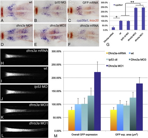
Dhrs3a functions as a regulator of RA signaling. (A?F) RNA in situ hybridizations on 5 somite stage embryos in dorsal view with anterior to the left, showing cyp26a1 expression (blue) in the anterior trunk. Red staining is erg2b in rhombomere 5 and aldh1a2 in the anterior paraxial mesoderm: (A) Uninjected control embryo; (B) control embryo injected with 5 ng tp53MO; (C) control embryo injected with 300 pg GFP mRNA; (D) embryo injected with 5 ng dhrs3a MO1 and 5 ng tp53 MO; (E) embryo injected with 5 ng dhrs3a MO3 and 5 ng tp53 MO; (F) embryo injected with 300 pg dhrs3a mRNA; (G) quantification of cyp26a1 expression by qPCR. (H?L) projections of confocal images through the spinal cord of live Tg(12XRARE-ef1a:gfp)sk71 transgenic embryos at 48 hpf. The anterior limit of GFP expression corresponds to the rhombomere 6/7 boundary: (H) dhrs3a mRNA-injected embryo; (I) uninjected control; (J) control tp53 MO-injected embryo; (K) dhrs3a MO3 plus tp53 MO-injected embryo; (L) dhrs3a MO1 plus tp53 MO-injected embryo. (M) quantification of live Tg(12XRARE-ef1a:gfp)sk71 signal in n = 6 injected with 300 pg dhrs3a mRNA, n = 6 uninjected, n = 6 tp53MO (5 ng/emb) injected, n = 6 tp53 + dhrs3a MO3 (5 ng/emb each) injected and n = 6 tp53 + dhrs3a MO1 (5 ng/emb each) injected embryos. ?Overall GFP expression? is the sum of pixel intensities above background threshold; ?GFP expression volume? is the total number of pixels in the confocal stack above background threshold. Values are normalized to the uninjected control. NN (ANOVA; the Newman?Kuels test) p < 0.005. N (ANOVA; the Newman?Kuels test) p < 0.05. There is no significant difference on the GFP expression volume or overall signal between wt and tp53. + (Student′s t-test) p < 0.05. dhrs3a mRNA injected embryos were normalized to control mRNA injected embryos. |
| Genes: | |
|---|---|
| Fish: | |
| Knockdown Reagents: | |
| Anatomical Terms: | |
| Stage Range: | 5-9 somites to Long-pec |
Reprinted from Developmental Biology, 338(1), Feng, L., Hernandez, R.E., Waxman, J.S., Yelon, D., and Moens, C.B., Dhrs3a regulates retinoic acid biosynthesis through a feedback inhibition mechanism, 1-14, Copyright (2010) with permission from Elsevier. Full text @ Dev. Biol.