Fig. S1
|
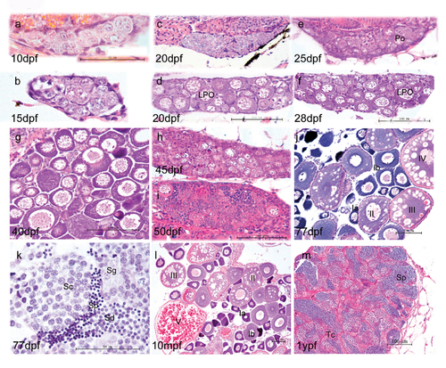
Morphological development of zebrafish gonads. At 10 dpf and 15 dpf (a, b), the majority of gonad cells are different types of germ cells, surrounded by basophilic somatic cells. In the subsequent juvenile period, some gonads show fast development with more meiotic germ cells and more condensed late perinucleolar oocytes (LPO) (d, f). The other gonads show more stromal cells and fewer LPO (c, e). By 40 dpf, most fish have undergone sex differentiation (g), although a few individuals still undergo ovary-testis transformation at 45 dpf (h) and 50 dpf (i). At 77 dpf, fish complete sex differentiation (j, k). j and l show the mature ovary at 77 dpf and 10 month postfertilization (mpf). As the functional unit of female ovary, follicles develop by a continuous production of preovulatory follicles and follicles of each stage (primordial, transitional, primary, activated primary, antral follicle). Adult ovaries show various stages of oogenesis: stage Ia, Ib, II, III, and IV oocytes. k and m show mature testis at 77 days post fertilization and 1 year post fertilization, respectively. The tubular compartment or testicular cord (Tc) is composed of clusters of germ cells enclosed by a layer of Sertoli cells. K and M show germ cells at various stages of spermatogenesis. Key: Spermatogonia (Sg), spermatocytes (Sc), spermatids (Sd) and sperm (Sp). Scale bar =50 μm (a,b,l) and 100 μm (c,d,e,f,g,h,I,m,n). |

