
Fig. S1
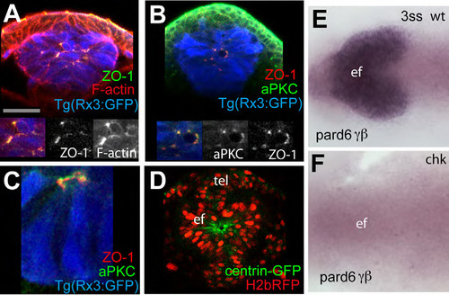
(A-C) 4ss Tg(rx3:GFP) embryos immunostained as detailed in the panels [ZO-1 (green in A, red in B and C), F-actin (red, A) and aPKC (green in B)]. Inset boxes showing F-actin and aPKC, and ZO- 1 and aPKC colocalise in the eye field at 4ss. aPKC can sometimes localise apically to ZO-1 (C). (D) View of the eyefield of a wildtype embryo injected with mRNA for H2b-RFP to label the nuclei and centrin-GFP to label centrosomes, showing accumulation of this marker at the centre of the eye field. White dotted lines in (D) outline the eye field. tel: telencephalon; ef: eye field. (E-F) in situ hybridization to detect pard6γβ in the eye field of wildtype (E) and rx3chk mutant embryos (F). |
Reprinted from Developmental Cell, 27(3), Ivanovitch, K., Cavodeassi, F., and Wilson, S.W., Precocious acquisition of neuroepithelial character in the eye field underlies the onset of eye morphogenesis, 293-305, Copyright (2013) with permission from Elsevier. Full text @ Dev. Cell