
FIGURE
Fig. S3
Fig. S3
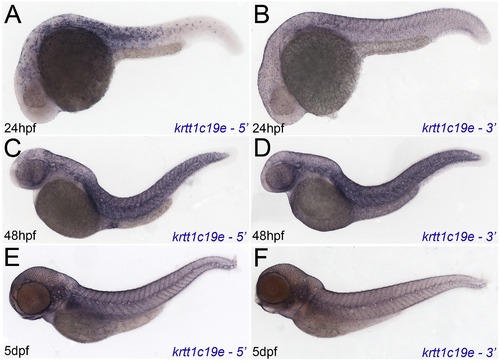
Independent in situ probes confirm krtt1c19e expression. Micrographs of 24hpf (A, B), 48hpf (C, D) and 5 dpf (E, F) embryos hybridised with 52 krtt1c19e (A, C, E) and 32 krtt1c19e (B, D, F) in situ probes. Whilst sensitivity was reduced, in particular at 24hpf, expression in the epidermis was identical to that seen with the full length probe. |
Expression Data
Expression Detail
Antibody Labeling
Phenotype Data
Phenotype Detail
Acknowledgments
This image is the copyrighted work of the attributed author or publisher, and
ZFIN has permission only to display this image to its users.
Additional permissions should be obtained from the applicable author or publisher of the image.
Full text @ PLoS One