
Fig. 10
- ID
- ZDB-FIG-140324-42
- Publication
- Weber et al., 2013 - Characterization of light lesion paradigms and optical coherence tomography as tools to study adult retina regeneration in zebrafish
- Other Figures
- All Figure Page
- Back to All Figure Page
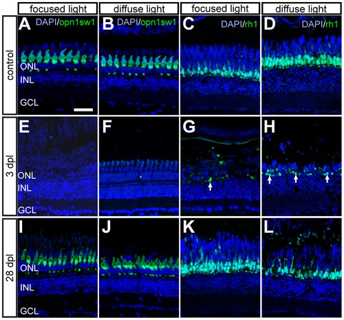
Time course of GFP+ photoreceptors after light lesions. UV cones (left panel) and rods (right panel) are labelled by the opn1sw1:GFP and rh1:GFP reporter line, respectively. A, B: Untreated control showing continuous mosaic pattern of UV cones. C, D: Untreated control showing the continuous rod layer in the ONL. E, F: Loss of all GFP+ cones 3 days after focused and diffuse light lesions. G, H: Partial depletion of rods 3 dpl after focused and diffuse light lesions. Examples of remaining rods are indicated by arrows. I, J: Regenerated UV cones in the ONL at 28 days after focused and diffuse light lesions. K, L: GFP+ rods regenerate in the appropriate layer after focused and diffuse light lesion. Scale bars represent 20 μm. |