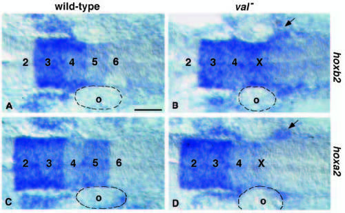
Paralogue group 2 gene expression in wild-type (A,C) and val- (B,D) embryos at the 20s stage reveals a population of hox expressing cranial neural crest migrating posterior to the otic vesicle. In wild-type embryos (A), hoxb2 is expressed in r3, r4 and r5 and in cranial neural crest deriving from r4 and migrating anterior to the otic vesicle (o) into the 2nd branchial arch. In val- embryos (B) the otic vesicle (o) is reduced in size; in addition to the normal population of cranial neural crest migrating anterior of the vesicle, there is a subpopulation possibly deriving from rX, which expresses hoxb2 but migrates posterior to the otic vesicle toward the 3rd arch (arrow). In wild-type embryos (C) hoxa2 is expressed in r2, r3, r4 and r5, and in cranial neural crest migrating into the 2nd and 3rd branchial arches. In val- embryos (D) expression levels are reduced in rX, however there are increased expression levels in neural crest migrating into the 3rd branchial arch (arrow). Scale bar, 50 μm.
|

