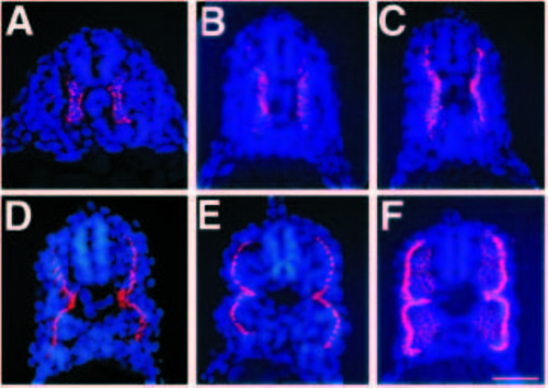
Adaxial cells migrate radially away from the notochord, between other somitic cells. In the caudal trunk, migration occurs over approximately 5 hours. Transverse sections through the caudal trunk (somites 14-17) were immunolabeled with F59 to mark adaxial cells and counter stained with Hoechst to reveal the nuclei (A-F). (A) 17 h (16 somites) embryo. F59 immunoreactivity is present only in adaxial cells, the labeling is perinuclear. (B) 18.5 h (19 somites) embryo. The adaxial cells have begun to move dorsally and ventrally; this is approximately the time that these cells are elongating in the anterior posterior dimension. (C) 20.5 h (23 somites) embryo. The adaxial cell population now extends almost the full dorso ventral extent of the myotome. (D) 21.5 h (25 somites) embryo. Radial migration of the adaxial cells has begun. (E) 23 h (28 somites) embryo. Most of the adaxial cells have reached the lateral surface. (F) 24 h embryo. The adaxial cells are now lateral. A subset of adaxial cells remains apposed to the notochord, generating an hourglass shape to the layer of superficial muscle cells. The deep cells of the myotome are now beginning to express the F59 epitope weakly. In these transverse sections, dorsal is up. Scale bar, 50 μm.
|

