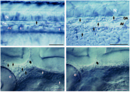
(A,B) Live embryos at 36 hours. As in wild type (A), the clo mutant (B) has blood vessels in the trunk. A shows the central vessels in the mid-trunk region; B shows the central vessels more caudally where they are easier to photograph in the mutant. Arrows point to endothelial cells. The dorsal aorta (DA) sits between the notochord (N) and the axial vein (AV). (Cells fill the trunk vessels in wild-type and mutant embryos but because they don?t circulate in the mutants, they are more clearly distinguished.) In the head, at 28 hours (which is after the initiation of the heart beat but before circulation is established in the head; Stainier et al., 1993), blood vessels are clearly seen in wild-type (C) but not in clo (D). Straight arrows point to endothelial cells; curved arrow points to the midbrain-hindbrain boundary. We also examined plastic sections throughout the head region and confirmed the Nomarski optics observations. At earlier times (and also here at 28 hours), the head mesoderm in clo appears more condensed than in wild-type, suggesting that the migrating angioblasts that normally form part of the loose head mesenchyme may in fact be missing. e, eye. Scale bars, 50 μm.
|
