
FIGURE
Fig. 3
- ID
- ZDB-FIG-140228-12
- Publication
- Round et al., 2014 - Slitrk gene duplication and expression in the developing zebrafish nervous system
- Other Figures
- All Figure Page
- Back to All Figure Page
Fig. 3
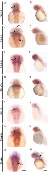
A-N Slitrk expression at 48 hr postfertilization (hpf) is observed in the developing nervous system. In situ hybridization performed on whole-mount zebrafish embryos at 48 hpf demonstrates differential expression patterns throughout the developing brain of seven different slitrks. A dorsal view of the head (left) and lateral view of the whole embryo (right) are provided for each slitrk gene. M, midbrain; NE, neuroepithelium; OV, otic vesicle; PG, pineal gland; RH, rhombomeres of the hindbrain. Scale bars, 50 μm. |
Expression Data
| Genes: | |
|---|---|
| Fish: | |
| Anatomical Terms: | |
| Stage: | Long-pec |
Expression Detail
Antibody Labeling
Phenotype Data
Phenotype Detail
Acknowledgments
This image is the copyrighted work of the attributed author or publisher, and
ZFIN has permission only to display this image to its users.
Additional permissions should be obtained from the applicable author or publisher of the image.
Full text @ Dev. Dyn.