
Fig. 9
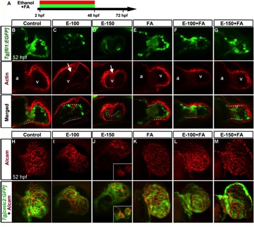
Folic acid supplementation during chronic ethanol exposure (2?48 hpf) rescues ethanol-induced endocardial cushion and chamber formation defects. A: Schematic diagram showing the timing of ethanol and FA exposure. B?G: Endocardial cushion formation was restored in ethanol-exposed, FA co-supplemented embryos. Stained Tg[fli1:EGFP] embryos showed clustering of GFP- and F-actin-positive cells (white dashed line) in the control, FA-treated, and ethanol+FA-cotreated embryos. Ethanol-treated embryos exhibited no clustering of GFP- and F-actin-positive cells at the AV boundary. White arrow, defective wall in the ventricle. a, atrium; v, ventricle. Atrium in the ethanol-treated embryos cannot be seen in this view. H?M: Ethanol-induced ventricular chamber defects were rescued in the FA-cosupplemented embryos. Alcam-stained Tg[cmlc2:GFP] embryos showed a similar pattern of cardiomyocyte distribution in the ventricular wall of control, FA-treated, and ethanol+FA-cotreated embryos; cardiomyocyte sizes and shapes were variable throughout the ventricular wall in the ethanol-treated embryos. Inset (J) in 150 mM ethanol showed cardia bifida. Anterior to the top. |