
Fig. S5
- ID
- ZDB-FIG-130826-25
- Publication
- Hollenbach et al., 2013 - Different Regulation of Physiological and Tumor Angiogenesis in Zebrafish by Protein Kinase D1 (PKD1)
- Other Figures
- All Figure Page
- Back to All Figure Page
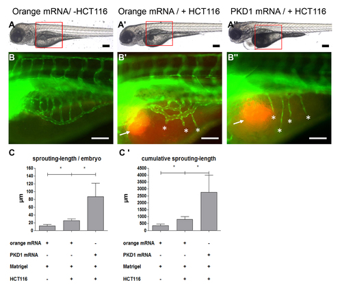
Increased tumor angiogenesis in tg(fli1:EGFP) zebrafish embryos overexpressing PKD1. Overall morphology of orange-mRNA - injected (100 pg) or PKD1 mRNA - injected (100 pg) 96 hpf embryos. At 48 hpf 1?4 nl of Matrigel (A, B) or Matrigel/HCT116 solution (A2, A3, B2, B3) was injected in the perivitelline space. Red boxes indicate regions of pictures shown in (B?B3). B?B3, HCT116 induced tumor angiogenesis as indicated by sprouting of subintestinal venous plexus (SIV) was analyzed at 96 hpf in tg(fli1:EGFP) embryos. Injection of HCT116 tumor cells (labeled with VybrantDil in red, arrow) led to a strong formation of ectopic blood vessels originated from the SIV (asterisks). In PKD1 overexpressing embryos ectopic blood vessel formation was further enhanced. C?C2, Quantification of sprouting length per embryo (C) and cumulative sprouting length (C2) with S.D. of at least 30 embryos per group. For cumulative sprouting-length all sprouts of the same number of embryos per group were summed, (C2) represents means of three independent experiments with S.D. Black scale bars: 300 μm; white scale bars: 100 μm. *P<0.05, **P<0.01, ***P<0.001. |