
Fig. 1
- ID
- ZDB-FIG-130808-62
- Publication
- Duval et al., 2013 - Longitudinal fluorescent observation of retinal degeneration and regeneration in zebrafish using fundus lens imaging
- Other Figures
- All Figure Page
- Back to All Figure Page
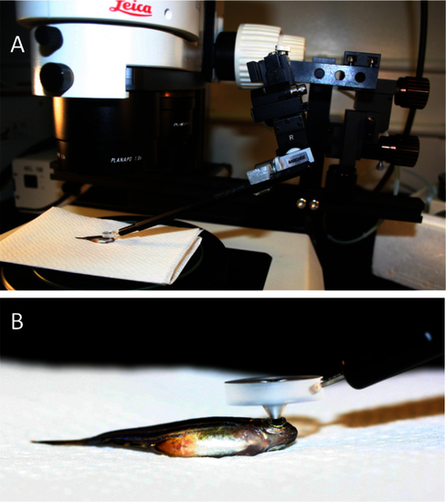
Mounting of a custom fundus lens on a fluorescent stereomicroscope allows characterization of individual photoreceptor cells in vivo. A: An adult anaesthetized zebrafish is shown, positioned on its flank under the objective lens such that its pupil is in the centre of the field-of-view. The fundus lens is positioned in the light path, centered above and touching the pupil (detailed in B). A micromanipulator (right side of image) allows precise and constant positioning of the fundus lens but is not required. B: A view of the custom fundus lens in position for viewing, positioned on the fish eye. The ventral side of the fish is in view. |