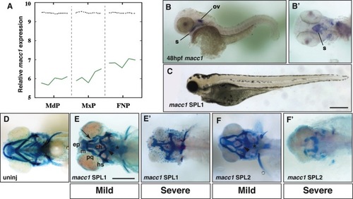
Analysis of macc1 expression and function in the larval zebrafish head. A: Microarray analysis of Macc1 expression in the embryonic mouse face. Expression increases in the mandibular (MdP), maxillary (MxP), and frontonasal (FNP) prominences between E10.5 to E12.5. The x-axis represents the five time points for each prominence. The y-axis shows the relative expression level on a log2 scale, i.e., every integer represents a doubling in the level of expression from the preceding number. The dashed black line indicates the average expression level for all probes. B: In situ hybridization for macc1 at 48hpf. Lateral view shows macc1 expression in the otic vesicle (ov) and developing mouth (s). B′: Ventral view after removal of the yolk to more clearly show macc1 expression in the roof of the stomodeum (s). C: Lateral view of 5dpf zebrafish after injection of 7.5 ng macc1 SPL1. The head and eyes of macc1 morphants are hypoplastic. D: Ventral view of the craniofacial skeleton of an uninjected larva at 5dpf. E: Ventral view of mild phenotypes associated with injection of 5 ng macc1 SPL1 Morpholino. E′: Ventral view of severe phenotypes associated with injection of 10 ng SPL1 macc1 Morpholino. F: Ventral view of mild phenotypes associated with injection of 10 ng macc1 SPL2 Morpholino. F′: Ventral view of severe phenotypes associated with injection of 20 ng SPL2 macc1 Morpholino. Defects in the ceratobranchials (asterisk), the ceratohyals (arrowhead), and Meckel′s and palatoquadrate cartilages resulting in a recessed lower jaw (arrow) are indicated. Other abbreviations as in Figure 1.
|
