
Fig. S1
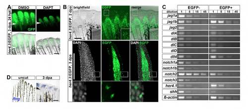
Expression of a transgenic reporter for Notch signaling and Notch receptors and ligands during fin regeneration. (A) In her4.3:EGFP transgenic regenerates treated with DAPT for 24 hours (24 hpt), EGFP fluorescence is reduced relative to DMSOtreated fins (n=3/3). (B) EGFP fluorescence in her4.3:EGFP transgenic regenerates is detectable in a few scattered cells in the wound epidermis. Top panel: whole-mount image of regenerates at 2 dpa. Bottom panel: confocal images of longitudinal sections of regenerates stained with GFP antibody and DAPI at 3 dpa. (C) Semi-quantitative PCR of the indicated genes on serial dilutions of cDNA derived from the EGFP-positive and EGFP-negative cellular pools of FACS-sorted her4.3:GFP regenerates at 3 dpa. (D) lunatic fringe (lfng) expression, as detected by whole-mount in situ hybridization, is upregulated in the regenerating fin at 3 dpa relative to uncut fins. Arrowheads indicate amputation plane. Scale bars: 200 μm. |