
Fig. S1
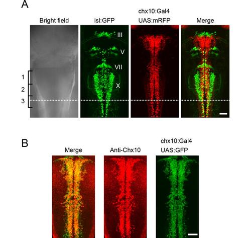
Hindbrain V2a neurons (A), A comparison of V2a neurons and cranial motoneurons in the hindbrain. Triple transgenic fish of Tg[chx10:Gal4], Tg[UAS:mRFP] and Tg[isl1:GFP] at 3dpf. Dorsal views. Nuclei of cranial motoneurons are shown in Roman numerals. The caudal end of the vagal motoneurons (X) is located near the middle of the third muscle segment (dotted line). This was used as a landmark for defining the caudal end of the hindbrain in this study. (B), Immunostaining of Chx10 protein in a compound transgenic fish of Tg[chx10:Gal4] and Tg[UAS:GFP] at 2 dpf. Dorsal views. The distribution of Chx10-positive cells and GFP-positive cells mostly overlap. Scale bar, 50 μm. |